Innate Immune Cells as Regulators of Mucosal Homeostasis & Wound Healing
Neutrophil (PMN) infiltration of the intestinal mucosa and accumulation in the intestinal lumen is a hallmark of Inflammatory Bowel Diseases (IBD). While en masse migration of PMNs across epithelial layers is often associated with epithelial injury, the function of transmigrated PMNs in the intestinal lumen, their interactions with luminal epithelial receptors and the consequent effects on epithelial homeostasis is unclear. Thus, ongoing studies in our laboratory are aimed at elucidating the mechanisms governing PMN-induced epithelial injury and alterations in epithelial barrier function. Our recent findings identified ICAM-1 as an important mediator of PMN retention and accumulation in the intestinal lumen and regulator of epithelial permeability and mucosal healing. Furthermore, recent evidence suggests that the interactions between PMNs and resident macrophages (including perivascular and crypt macrophages, PVMs and RCMs, respectively) may contribute to trafficking of these cells and the regulation of intestinal homeostasis. Thus, we are interested in defining PMN tissue interactions with resident macrophages and luminal interactions with epithelial cells in models of IBD and mucosal injury.
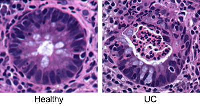
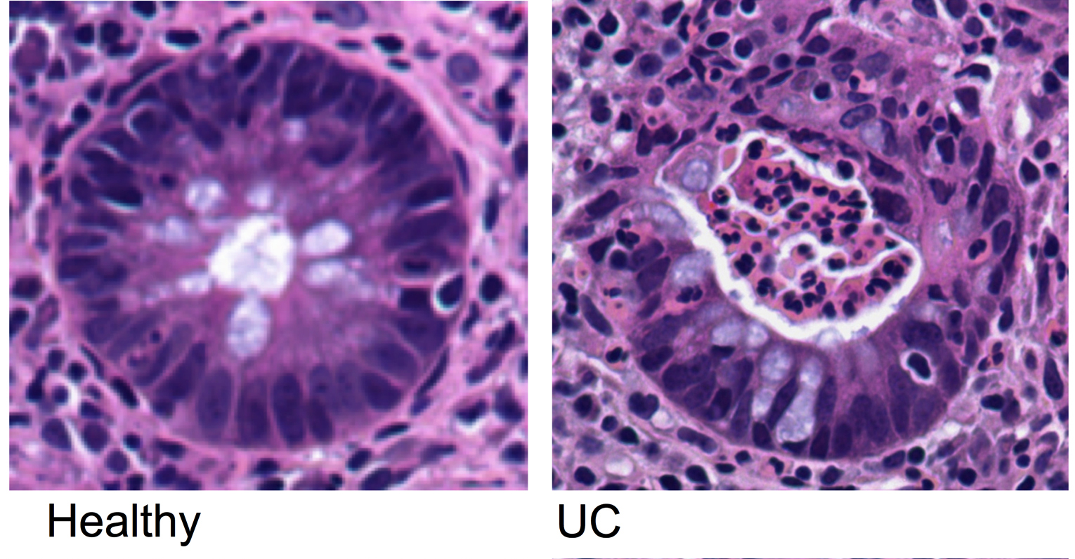
Mucosal epithelium from a healthy patient (left) showing intact columnar epithelial cells that form a tight barrier between the intestinal lumen and the surrounding tissue. Mucosal epithelium from a patient with ulcerative colitis (right) showing epithelial damage and luminal accumulation of migrated neutrophils, characteristic of crypt abscesses as seen in IBD.
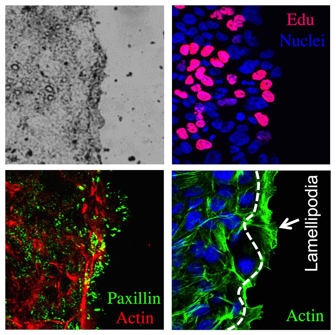
In vitro time-lapse imaging approaches to study wound closure using actin-eGFP epithelial cells.
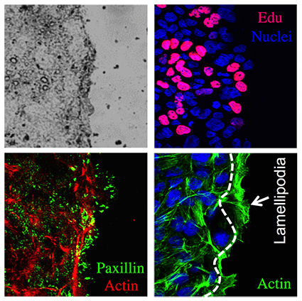
Two essential components of wound healing are epithelial cell proliferation and migration. We are interested in studying both processes under various conditions using live imaging and immunofluorescence staining approaches. From left to right are shown scratch-wounded epithelial monolayers (bright field illumination), proliferation of the leading edge cell (proliferating cells are red), increases in expression of the focal adhesion protein, paxillin (green), during cell spreading and extension of lamellipodia during cell migration (actin is green).